Зачетное задание по 1 блоку 2 семестра
advertisement
Зачетное задание по 1 блоку 2 семестра
Уже второй семестр я работаю с информацией о белке IHFA_ECOLI, название
которого можно перевести как «альфа-субъединица интеграционного фактора». Его
молярная масса 11353,79164. AC : P0A6X7; P06984; ID IHFA_ECOLI.
Он представляет собой одну из двух субъединиц фактора интеграции в хозяина,
специфического ДНК-связывающего белка, участвующего в генетической рекомбинации,
а также в транскрипционном и трансляционном контроле. IHFА также играет важную
роль в лизогенном жизненном цикле лямбда-фага, котороя необходима нетолько для
рекомбинантной реакции, вставляющей ДНК лямбды в хромосому Е.Сoli, но и для синтеза
int и cI репрессоров, двух фаговых белков, необходимых для внедрения и
репрессирования ДНК соответственно.
IHF образован двумя субъединицами IHFA и IHFB. Их длины соответственно 99 и
94 а.к. остатка. IHF образует комплекс с ДНК (т.е. в данном случае ее можно считать
органическим лигандом.) из неорганических лигандов только ионы кадмия и вода.
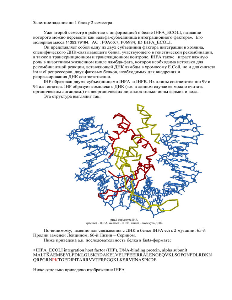
Эта структура выглядит так:
рис.1 структура IHF.
красный – IHFA, желтый – IHFB, синий – молекула ДНК.
По-видимому, именно для связывания с ДНК в белке IHFA есть 2 мутации: 65-й
Пролин заменен Лейцином, 66-й Лизин – Серином.
Ниже приведена а.к. последовательность белка в fasta-формате:
>IHFA_ECOLI integration host factor (IHF), DNA-binding protein, alpha subunit
MALTKAEMSEYLFDKLGLSKRDAKELVELFFEEIRRALENGEQVKLSGFGNFDLRDKN
QRPGRNPKTGEDIPITARRVVTFRPGQKLKSRVENASPKDE
Ниже отдельно приведено изображение IHFA
рис.2 третичная структура белка IHFA
Белок выделен из организма E.Coli, с ДНК которой он связывается.
Ссылки на записи о моем белке в различных базах данных:
PDB , UniProt, SRS.
Ниже приведены аннотации к некоторым статьям о моем белке, которые показались
мне в большей степени, чем остальные посвященными именно белку IHFA_ECOLI:
1."Sequence of the Escherichia coli pheST operon and identification of the himA gene."
Mechulman Y., Fayat G., Blanquet S.
J. Bacteriol. 163:787-791(1985)
The complete nucleotide sequence of the Escherichia coli pheST operon coding for the two
subunits of phenylalanyl-tRNA synthetase (an alpha 2 beta 2-type enzyme) has been determined.
Another open reading frame (prp) was revealed downstream from pheT which was identified as
himA, the gene for the alpha subunit of the integration host factor.
2. "Crystal structure of an IHF-DNA complex: a protein-induced DNA U-turn."
Rice P.A., Yang S., Mizuuchi K., Nash H.A.
Cell 87:1295-1306(1996)
Integration host factor (IHF) is a small heterodimeric protein that specifically binds to
DNA and functions as an architectural factor in many cellular processes in prokaryotes. Here, we
report the crystal structure of IHF complexed with 35 bp of DNA. The DNA is wrapped around
the protein and bent by >160 degrees, thus reversing the direction of the helix axis within a very
short distance. Much of the bending occurs at two large kinks where the base stacking is
interrupted by intercalation of a proline residue. IHF contacts the DNA exclusively via the
phosphodiester backbone and the minor groove and relies heavily on indirect readout to
recognize its binding sequence. One such readout involves a six-base A tract, providing evidence
for the importance of a narrow minor groove.
3. "The isolation and characterization of mutants of the integration host factor (IHF) of
Escherichia coli with altered, expanded DNA-binding specificities."
Lee E.C., Hales L.M., Gumport R.I., Gardner J.F.
EMBO J. 11:305-313(1992)
The integration host factor (IHF) of Escherichia coli is a small, basic protein that is
required for lambda site-specific recombination and a variety of cellular processes. It is
composed of two subunits, alpha and beta, that are encoded by the himA and hip (himD) genes,
respectively. IHF is a sequence-specific DNA-binding protein and bends the DNA when it binds.
We have used the bacteriophage P22-based challenge phage selection to isolate suppressor
mutants with altered, expanded DNA binding specificities. The suppressors were isolated by
selecting mutants that recognize variants of the phage lambda H'IHF recognition site. Two of the
mutants recognize both the wild-type and a single variant site and contain amino acid
substitutions at positions 64 (Pro to Leu) or 65 (Lys to Ser) of the alpha subunit. These
substitutions are in a region of the protein that is predicted to contain a flexible arm that interacts
with DNA. Three other mutants, which recognize the wild-type and a different variant site,
contain amino acid substitutions at position 44 (Glu to Lys, Val or Gly) of the beta subunit.
These substitutions are in the middle of a predicted beta-strand of the subunit. We discuss the
possible mechanisms of suppression by the mutants in terms of a model of the IHF-DNA
complex proposed by Yang and Nash [Cell, 57, 869-880 (1989)].
